Neutral Red Uptake

The Normal Human Epidermal Keratinocytes (NHEKs) Cytotoxicity Assay using a Neutral Red Uptake (NRU) viability endpoint is a 96-well cytotoxicity assay to assess the toxicity potential of a test material. The assay utilizes primary NHEK cells and may be used to predict either human lethal serum concentrations or in vivo rodent LD50 starting doses for acute oral systemic toxicity. Cytotoxicity caused by the test article is measured by a concentration-dependent reduction in neutral red uptake (NRU) by the cells after exposure to a test material.

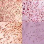
Healthy mammalian cells, when maintained in culture, continuously proliferate. A toxic chemical, regardless of site or mechanism of action, will interfere with this process and result in a reduction in viable cells relative to untreated controls. The viability of the cells can be assessed using a neutral red uptake endpoint. A decrease in the uptake of neutral red dye in treated cell cultures following a test chemical exposure is used to determine relative toxicity.

Quick Facts

Assay Model: NHEK cells seeded in a 96-well plate

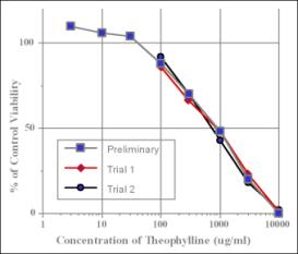
Endpoint: IC50 (the concentration of the test material that causes a 50% decrease in viability, relative to solvent control)


Dose Response Curves - Viability Assessed by NRU in NHEK Cells

Dose Response Curves – Viability Assessed by NRU in NHEK Cells

NHEK Cytotoxicity Neutral Red Uptake Applications

Test Material Compatibility

The 96-well plate based cytotoxicity assays using NRU are best suited for water-soluble materials; however, test materials may be dissolved in intermediate solvents so that they are compatible with the test system. At IIVS, a solubility test is performed to find the most suitable solvent when the solubility of a test chemical is unknown or unavailable.

As a Prediction of Systemic Toxicity

The NHEK Cytotoxicity Assay using a NRU viability endpoint can be used to estimate human lethal serum concentrations, according to a prediction model developed in the Multicentre Evaluation of In Vitro Cytotoxicity (MEIC) program (Part I of this program presents the methodology of 68 in vitro toxicity assays used to test the first 30 chemicals. (MEIC Evaluation of Acute Systemic Toxicity: Part I: C. Clemedson et al, 1996, ATLA 24, 249-272)).

The NHEK Cytotoxicity Assay using a NRU viability endpoint can also be used to estimate starting doses for acute oral toxicity testing in rodents (the methods were initially proposed by ZEBET based upon the correlation of the in vitro cytotoxicity endpoint IC50 and the in vivo LD50 values for 347 chemicals as presented in the Registry of Cytotoxicity (Halle, 2003)).

As a Prediction of Ocular Irritancy

Since the target cells (NHEKs) are epithelial in origin, NHEK Cytotoxicity Assay using a NRU viability endpoint may be used to predict ocular irritation for aqueous soluble materials. This approach has been used to determine the relative ocular irritation potential of surfactants and preservatives, and may be well suited for evaluating relative irritancy potentials of ingredients in ophthalmic solutions in a product development setting.

Specialized Protocols

Test article concentrations may be adjusted as necessary to accommodate specific physical characteristics or client needs. Specialized protocols may be prepared as requested through consultation with the Study Director.

Validation

In 2007, ICCVAM recommended both in vitro test methods (3T3 NRU and NHEK NRU) as reduction alternatives to estimate the starting dose in the Up-and-Down Procedure and Fixed Dose Procedure for assessing acute oral systemic toxicity. Recommendations were accepted by U.S. Federal agencies, and OECD Guidance Document 129 for implementation of the test methods was published in 2010.

Step-by-Step

  1. Seed
  2. Dose
  3. Test Article Removal & Rinsing
  4. Addition of Vital Dye
  5. Addition of Solvent
  6. 96-well Plate Reading
Step 1: Seed

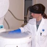
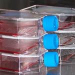
The cells to be used for the 96-well assays are stored in a liquid nitrogen freezer. When ready for use, the cells are thawed, resuspended, and transferred to a culture flask, where they will grow and multiply until optimal conditions have been achieved.

Trypsin solution is added to the flask to remove the cells. The cells are resuspended in medium, counted, and “seeded” into 96-well plates using a multichannel pipet.

Cell storage
Cell amoule
Cell culture
96 well plate cultures
Step 2: Dose

The test material is serially diluted to make a range of doses.

The test article dilutions are added to the inner wells containing cells.

Each plate also contains 12 negative, or solvent, control wells which are used to determine % of control viability.

Serial dilution series
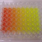
Dosed 96 well plate
Step 3: Test Article Removal & Rinsing

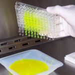
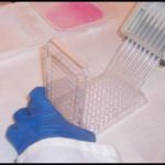
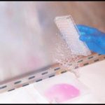
The test material is removed by decanting.

The plates are rinsed with a buffered saline solution to remove any residual test material.

Test material removal
Test material rinsing
Rinsate removal
Step 4: Addition of Vital Dye

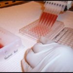
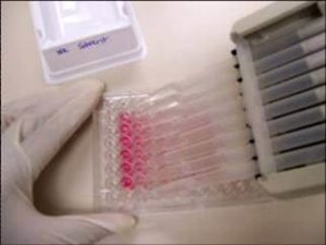
A solution of Neutral Red, a vital dye, is added to the 96-well plate. The plates are incubated at standard culture conditions to allow neutral red uptake by the cells.

Addition of Neutral Red for 3T3 Phototoxicity Assay
Neutral red uptake
Step 5: Addition of Solvent
Solvent addition

Solvent addition

After the 3 hour neutral red incubation, excess neutral red is decanted and solvent is added to all wells.

The solvent extracts the neutral red dye contained within the cells.

Step 6: 96-well Plate Reading
Optical Density Determination

Optical Density Determination

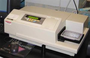
The 96-well plates are placed on a plate shaker to fully extract the neutral red and evenly distribute the dye in each well.

A spectrometer measures the absorbance of each sample at a specific wavelength.

The absorbance values (optical density) are then used to determine the viability of each well by comparing the optical density of the each test material treated well compared the negative (or solvent ) control wells.

Assays of Interest