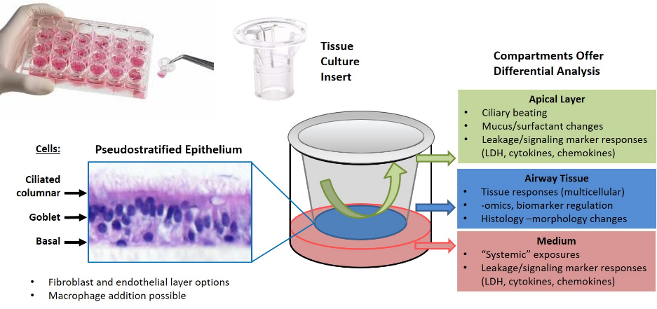
Recent technology advances has allowed the development of reconstructed airway epithelium. Primary cells obtained from donor tissue are expanded, seeded onto porous membranes, and cultured at an Air-Liquid-Interface (ALI) into a pseudostratified mucociliary tissue that recapitulate key physiologic functions. The RHuA contain multiple cell types including ciliated columnar cells, mucus-producing Goblet cells, and basal cells. IIVS has strong ties to both MatTek, Inc. and Epithelix Sarl; manufacturers of high quality tissues, EpiAirway™ and MucilAir™, respectively. Available RHuA tissues can be generated from cells of nasal, tracheal, and bronchial origin and be of healthy or diseased origin. Cultures can extend to weeks and months and allow the observation of short term (e.g. changes in ciliary beat frequency; CBF) and long term events (e.g. Goblet cell hyperplasia; GCH). Recent additions to the portfolio of RHuA cultures grown at ALI include Epithlix’s SmallAir™ and MatTek’s EpiAlveolar™ – tissues modeling the small airways and alveolar regions of the lung, respectively. The tissues generate inflammatory cytokine responses and are unique in that they are a 3D multi-cellular model with distinct sampling compartments, including the airway space that allows inhalation-like exposures.

